
Odisha State Board CHSE Odisha Class 11 Biology Solutions Chapter 19 Excretory Products and Their Elimination Textbook Questions and Answers.
CHSE Odisha 11th Class Biology Chapter 19 Question Answer Excretory Products and Their Elimination
Excretory Products and Their Elimination Class 11 Questions and Answers CHSE Odisha
Very Short Answer Type Questions
Multiple Choices Questions
Question 1.
The organs of excretion in the cockroach are
(a) Flame cells
(b) Green gland
(c) Nephridia
(d) Malpighian tubules
Answer:
(d) Malpighian tubules
Question 2.
Birds eliminate their nitrogenous wastes in the form of
(a) ammonia
(b) urea
(c) uric acid
(d) amino acids
Answer:
(c) uric acid
Question 3.
Ornithine cycle occurs in
(a) liver
(b) kidney
(c) brain
(d) skin
Answer:
(a) liver
Question 4.
Ornithine cycles synthesises
(a) ammonia
(b) urea
(c) uric acid
(d) xanthine
Answer:
(b) urea
Question 5.
What is the main nitrogenous waste product in reptile?
(a) ammonia
(b) urea
(c) uric acid
(d) hippuric acid
Answer:
(c) uric acid
Question 6.
Urea is formed from the breakdown of
(a) carbohydrates
(b) proteins
(c) fats
(d) nucleic acids
Answer:
(b) proteins
Question 7.
In man, uric acid is formed from the break down of
(a) carbonydrates
(b) proteins
(c) fats
(d) nucleic acids
Answer:
(d) nucleic acids
Question 8.
Desert mammals have in their nephrons.
(a) long loops of Henle
(b) long proximal convoluted tubules
(c) long distal convolutions
(d) long collecting ducts
Answer:
(a) long loops of Henle
Question 9.
ADH exercises its action on part of the nephron.
(a) PCT
(b) Henle’s loop
(c) DCT
(d) glomerulus
Answer:
(c) DCT
Question 10.
Most aquatic animals are
(a) ammonotelic
(b) ureotelic
(c) ureotelic
(d) aminotelic
Answer:
(a) ammonotelic
Question 11.
What is diabetes insipidus due to?
(a) loss of glucose by the urine
(b) deficiency of ADH
(c) deficiency of insulin
(d) All of the above
Answer:
(b) deficiency of ADH
Question 12.
Which hormone is secreted from juxtaglomerular apparatus of kidney?
(a) renin
(b) angiotensin
(c) adrenalin
(d) calcitrol
Answer:
(a) renin
Question 13.
Reabsorption of sodium and chloride occurs in
(a) Ascending limb of Henle’s loop
(b) Proximal convoluted tubule
(c) Descending limb of Henle’s loop
(d) Distal convoluted tubule
Answer:
(b) Proximal convoluted tubule
Question 14.
Human kidney is
(a) pronephric
(b) mesonephric
(c) metanephric
(d) opisthonephric
Answer:
(b) mesonephric
Question 15.
Longer loop of Henle is meant primarily for increased absorption of
(a) glucose
(b) water
(c) potassium
(d) amino acids
Answer:
(b) water
Short Answer Type Questions
Write briefly on the following (within 50 words each)
Question 1.
Ammonotelism
Answer:
Ammonia is the most toxic form of nitrogenous waste, it requires large amount of water for its elimination. The organisms that excrete ammonia are called ammonotelic and this process of eliminating ammonia is known as ammonotelism. Examples of ammonotelic animals are bony fishes, aquatic amphibians and aquatic insects. Since, ammonia, is readily soluble in water and is generally excreted by diffusion across body surfaces or through gill surfaces (in fish) as ammonium ions the ammonotelism is commonly found in aquatic animals.
Question 2.
Ureotelism
Answer:
The process of excreting urea is called ureotelism. Animals, which do not live in high abundance of water convert ammonia produced in the body into urea (in the liver) and release it into the blood, which is filtered and excreted out as urine by the kidneys.
Examples of ureotelic animals are mammals, many terrestrial amphibians and marine fishes.
Question 3.
Uricotelism
Answer:
The process of excreting uric acid is called uricotelism. Uric acid, being the least toxic nitrogenous waste can be removed with a minimum loss of water from the animal body.
Thus, it is excreted in the form of pellet or paste (i.e., semisolid form). Normally, the animals which live in desert exhibit uricotelism.
Examples of uricotelic animals are reptiles, birds, land snails and insects.
Question 4.
Role of the Malpighian tubule in the excretion in cockroach.
Answer:
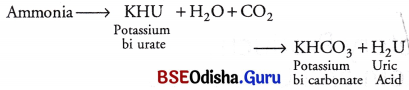
Malpighian tubules are principle excretory organs of a cockroach. These tubules consists of two distinct regions, i. e. the distal half (secretory) and the proximal portion (absorptive in function).
The distal region is alkaline, concerned with the secretion of urates of potassium and sodium extracted from the surrounding haemolymph.
The proximal region is acidic due to CO2 secretion.
The uric acid is therefore precipitated while sodium and potassium Eire reabsorbed as bicarbonates to be used again. The urine passes to ileum, excreted via rectum along with the faeces where excess water is absorbed from them.
Question 5.
Structure of the mammalian nephron
Answer:
The LS of a mammalian kidney shows differentiation into outer cortex and inner medulla.
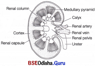
LS of kidney
Inside the kidney, the ureter is expanded as a funnel-shaped cavity called pelvis. The free end of pelvis has number of cup-like cavities called major calyces (sing, calyx) and minor calyces.
Medulla projects into the calyces as conical processes, called renal pyramids or medullary pyramids. The tip of pyramids are called renal papillae. The cortex spreads in between medullary pyramids as renal columns called columns of Bertini.
Microscopic Structure:
Each kidney is composed of numerous (nearly one million) complex tubular structures called nephrons. These are the functional units of kidney.
Structure of Nephron or Uriniferous Tubule
Each nephron consists of two parts, i.e., the Malpighian body or renal corpuscle and the renal tubule.
Malpighian Body or Renal Corpuscle Glomerulus along with Bowman’s capsule is called the Malpighian body or renal corpuscle which filters out large solutes from the blood and delivers small solutes to the renal tubule for modification.

Malpighian body (renal corpuscle)
Question 6.
Ultrafiltration
Answer:
Ultrafiltration or Glomerular Filtration
The first step of urine formation is the filtration of blood. It is carried out by the glomerulus. That is why this step is called glomerular filtration.
Kidneys filter about 1100-1200 mL of blood per minute, which constitute roughly 1/5th of the blood pumped out by each ventricle of the heart in a minute.
The glomerular capillary blood pressure causes filtration of blood through three layers, i.e.,
- the endothelium of glomerular blood vessels.
- the epithelium of Bowman’s capsule.
- a basement membrane (present between the above mentioned two layers).
The podocytes (epithelial cells of Bowman’s capsule) are arranged in such a manner, so that some minute spaces called filtration slits or slit pores occur between them.

Diagram representing the path taken by fluid (glomerular filtrate) during its passage from the plasma in a glomerular capillary of the lumen of a renal capsule
Question 7.
Selective reabsorption
Answer:
Selective Reabsorption:
This is the second step in the formation of urine from filtrate. The urine released is 1.5 L as compared to the volume of the filtrate formed per day (180 L). It suggests that as much as 99% of the material in the filtrate is reabsorbed by the renal tubules. Thus, the process is called reabsorption. Depending upon the types of molecules being reabsorbed, movements into and out of epithelial cells in different segments of nephron occur either by passive transport or active transport.
These are described as follows
(i) Water and urea, are reabsorbed by passive transport (i.e., water is reabsorbed by osmosis and urea by simple diffusion).
(ii) Glucose and amino acids are reabsorbed by activi transport.
(iii) The reabsorption of Na+, occurs both by passive and active transport.
Question 8.
Henle’s loop
Answer:
Henle’s Loop:
Reabsorption in Henle’s loop is minimum, besides this, it plays an important role in maintaining the high osmolarity of medullary interstitial fluid. Two portions of Henle’s loop, play different role in osmoregulation. These are as follows
1. Descending Limb of Loop of Henle Water is reabsorbed here due to increasing osmolarity of interstitial fluid but, sodium and other electrolytes are not reabsorbed here. This concentrates the filtrate as it moves down.
2. Ascending Limb of Loop of Henle This segment is impermeable to water, but permeable to K+,Cl– and Na+ and partially permeable to urea. Thus, in the thick ascending limb of the loop of Henle Na+, K+, Mg2+ and Cl are reabsorbed.
Therefore, as the concentrated filtrate pass upward, it gets diluted due to the passing out of electrolytes to the medullary fluid of kidney
Question 9.
Counter current mechanism
Answer:
Kidney of higher vertebrates (such as mammals, birds and man) has the ability of absorbing more and more water from tubular fdtrate to conserve water and make the urine more concentrated.
This can be achieved by a special mechanism known as counter current mechanism. The basic concept and this mechanism is as follows
Question 10.
Kidney as an endocrine organ
Answer:
Kidney as an endocrine glands The mammalian kidney performs many endocrine functions in the body. The kidney produces three hormones, i.e.
(a) Erythropoietin a peptide hormone which controls erythrocyte production in the body.
(b) Calcitriol the final activation of vitamin-D to active hormone calcitriol occurs in the kidneys.
(c) Renin is the part of the Renin-Angiotensin- Aldosterone System (RAAS). It controls the formation of angiotensin that influences blood pressure and sodium balance in the body.
Kidneys also synthesise prostaglandins, which Eiffect many processes occuring in the kidneys.
Question 11.
Secretion
Answer:
It is also an important step in urine formation. Certain chemicals in the blood that are not removed by filtration . from the glomerular capillaries are removed by this process of tubular secretion.
Acid base
It helps in the maintenance of ionic and acid-base balance of body fluids by removing chemicals like foreign bodies, ions (K+ , H+ , NH+4) and molecules (medicines), etc., that are toxic at elevated levels.
Question 12.
Acid-base balance
Answer:
Acid base
It helps in the maintenance of ionic and acid-base balance of body fluids by removing chemicals like foreign bodies, ions (K+ , H+ , NH+4) and molecules (medicines), etc., that are toxic at elevated levels.
Question 13.
Role of liver in excretion
Answer:
Role of Liver:
It changes the decomposed haemoglobin of the worn out red blood corpuscles into bile pigments, i.e., bilirubin and biliverdin. These pigments pass into the alimentary canal with the bile for elimination in the faeces. The liver also excretes cholesterol, steroid hormones, certain vitamins and drugs via bile.
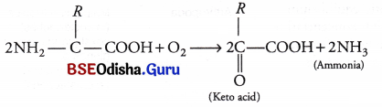
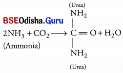
Liver deaminates the excess and unwanted amino acids, producing ammonia, which is quickly combined with CO2 to form urea in urea cycle or Ornithine cycle, which is further removed by the kidneys.
Question 14.
Orinithine cycle/Urea cycle
Answer:
Urea formation occurs in the liver by urea cycle or Ornithine or Kreb’s-Henseleit cycle. The excess amino acids in the body undergoes deamination in the liver and forms ammonia. Being highly toxic, ammonia is immediately converted to the less toxic urea by urea cycle. Urea cycle involves three amino acids, i.e. ornithine, citrulline and arginine.

Steps
(a) Ornithine reacts with ammonia and C02 forming citrulline and water.
(b) Citrulline combines with more ammonia forming arginine and water.
(c) Arginine then transforms into urea and ornithine in presence of enzyme arginase water.
Question 15.
Dialysis
Answer:
When the kidneys can no longer excrete water and ions at rates that maintain body balance of these substances nor they can excrete the wastes as fast as they are produces a technique called dialysis is used to cure kidney failure haemodialysis is carried out in which blood is filtered. Dialysis means separation of substances using a semi or a selectively permeable membrane.
Question 16.
Storage excretion in cockroach
Answer:
In cockroach, urate cells are found. These cells absorb and store uric acid throughout the life. This is called storage excretion as they remain stored in the cells of corpora adipose.
Question 17.
Renin-angiotensin system
Answer:
As blood pressure/glomerular blood flow /GFR decreases, the cells of the JGA release the enzyme renin.
Renin converts angiotensinogen in blood to Angiotensin I and Angiotensin II (active form). This mechanism is generally known as the Renin-angiotensin mechanism. Renin is the rate limiting factor in this system.
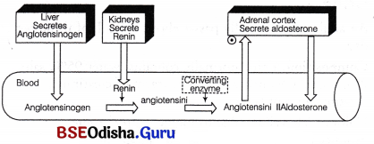
Summary of the renin angiotension aldosterone system
Question 18.
Obligatory water loss
Answer:
The minimal amount of fluid loss that can occur from the living body is known as the obligatory water loss.
Human kidney have the ability to produce a hyperosmotic urine that enables the body to survive without water for a long period. The human kidneys can produce a maximal urine concentration of 1400 m osmol/L., approx, five times the osmolarity of the blood plasma; which is 300 m osmol/L. Urea, sulfate, phosphate and other waste products and ions excreted each day amount to approximately 600 m osmol/L. The minimum volume of water in which the above quantity of solute can be dissolved and excreted in the urine is
= \(\frac{600 \mathrm{~m} \mathrm{Osmol} / \text { day }}{1400 \mathrm{~m} \mathrm{Osmol} / \mathrm{L}}\)
= 0.444 L/day
This volume of urine is referred to the ‘obligatory water loss’. The loss of this minimal volume of urine contributes to dehydration when a person goes without water for a long period.
G Explain the Following
Question 19.
Net filtration pressure
Answer:
Net Alteration pressure It is the pressure that helps to move filterate from the glomerulus into Bowman’s capsule. It is a combination of glomerular hydrostatic pressure, capsular hydrostatic pressure and osmotic colloid pressure.
Question 20.
Glomerular filtration rate
Answer:
Glomerular Filtration Rate:
The amount of the filtrate formed by the kidneys per minute is called Glomerular Filtration Rate (GFR). In a healthy person it was found approximately 125 mL /min, i. e., 180 L/day. GFR is regulated by one of the efficient mechanism carried out by Juxtaglomerular Apparatus (JGA).
JGA is a special sensitive region formed by cellular modifications in the distal convoluted tubule and the afferent arteriole at the location of their contact.
This apparatus includes
(i) granular juxtaglomerular cells in the afferent arteriole.
(ii) macula densa cells of DCT.
(iii) agranular lacis cells situated in between the above two.
Question 21.
Obligatory reabsorption of water
Answer:
Obligatory reabsorption of water Eighty five percent of the water reabsorption occurs irrespective of the body water balance and is called obligatory reabsorption of water. It takes place in proximal and distal tubules.
Question 22.
Role of ADH in water reabsorption
Answer:
Excessive loss of fluid from the body activates osmoreceptors present in blood vessels of hypothalamus, thus stimulating it to release ADH or vasopressin from the neurohypophysis. ADH release increases water reabsorption from DCT. An increase in body fluid volume switches off the osmoreceptors and suppresses the ADH release to complete the feedback. ADH also causes constrictory effects on blood vessels of glomerulus thus, increasing pressure for faster filtration.
Question 23.
Obligatory water loss
Answer:
The minimal amount of fluid loss that can occur from the living body is known as the obligatory water loss.
Human kidney have the ability to produce a hyperosmotic urine that enables the body to survive without water for a long period. The human kidneys can produce a maximal urine concentration of 1400 m osmol/L., approx, five times the osmolarity of the blood plasma; which is 300 m osmol/L. Urea, sulfate, phosphate and other waste products and ions excreted each day amount to approximately 600 m osmol/L. The minimum volume of water in which the above quantity of solute can be dissolved and excreted in the urine is
= \(\frac{600 \mathrm{~m} \mathrm{Osmol} / \text { day }}{1400 \mathrm{~m} \mathrm{Osmol} / \mathrm{L}}\)
= 0.444 L/day
This volume of urine is referred to the ‘obligatory water loss’. The loss of this minimal volume of urine contributes to dehydration when a person goes without water for a long period.
Question 24.
Functions of vasa recta
Answer:
Function of vasa recta
(i) Vasa recta plays an important role in counter-current mechanism.
(ii) Vasa recta also regulates the blood flow in kidney.
Differentiate between
Question 25.
Ammonotelism and Ureotelism
Answer:
Ammonotelism and Ureotelism
| Ammonotelism | Ureotelism |
| (a) The chief excretory waste product is ammonia. | The chief nitrogenous waste product is urea. |
| (b) Ammonia is highly soluble in water and is produced by deamination of amino acids. | Ureajs less soluble in water and is produced by urea cycle in liver via arginine. |
| (c) Ammonia waste is highly toxic and requires large quantity of water for elimination. | Urea is less toxic and does not require high abundance of water for elimination. |
| (d) The animals performing ammonotelism are called as ammonotelic. | The animals carrying out ureotelism are called ureotelic. |
| e.g. Protozoa, sponges, coelenterates, prawn, etc. | e.g. Mammals, earthworm, cartilaginous fish. |
Question 26.
Ureotelism and Uricotelism
Answer:
Ureotelism and Uricotelism
| Ureotelism | Uricotelism |
| The process of excreting urea is called ureo elism. | The process of excreting uric acid as waste product is called uricotelism. |
| Urea is less toxic than ammonia containing nitrogenous waste products. | Uric acid is the least toxic nitrogenous waste product. |
| Urea is formed in the liver by detoxification of ammonia. | Uric acid is formed mainly from the purines in liver cells. |
| It is characteristic excretory process of mammals, cartilaginous fishes and amphibians, etc. Such animals are called as ureotelic. | Uricotelism is characteristic of terrestrial reptiles insects and all birds. Such animals carrying out uricotelism are called uricotelic. |
Question 27.
Cortical nephron and juxamedullary nephron
Answer:
Cortical nephron and juxamedullary nephron
| Cortical nephron | Juxtamedullar neptron |
| They form 80% of the total nephron count. | These constitute 20% of total nephron count. |
| Cortical nephrons are smaller in size. | Juxtamedullary nephrons are larger in size. |
| The loop of Henle is too short, extends very little into the medulla. | The loop of Henle is very long and runs deep into the medulla. |
| Cortical nephrons lie in the renal cortex of kidney. | Juxtamedullary nephrons are found in the medulla of kidney. |
Question 28.
Selective reabsorption and secretion.
Answer:
Selective reabsorption and secretion.
| Tubular reabsorption | Tubular secretion |
| It involves the absorption of water and useful solutes from the glomerular filtrate into the blood. | It refers to the passage of waste materials from blood into the filtrate or nephrons. |
| It occurs by back diffusion and active transport. | It takes place only by active transport. |
| It occurs mostly in PCT. | It occurs mostly in DCT. |
| It does not occur in animals that lack glomerulus (e.g, marine fish and desert animals). | It is the only mode of excretion in animals that lack glomerulus. |
Question 29.
Haemodialysis and peritoneal dialysis
Answer:
Haemodialysis and peritoneal dialysis
| Haemodialysis | Peritoneal dialysis |
| Haemodialysis involves an artificial membrane in a dialyser machine. | Peritoneal dialysis uses the lining of the individual’s own abdominal cavity (peritoneum) as a dialysing membrane. |
| The process takes about 6-8 hours and is done atleast twice a week. | The peritoneal dialysis usually involves 4-6 exchanges of dialysing fluid per day. |
| It increases the risk of bloodstream related infections. | It increases the risk of an infection of peritoneum lining called peritonitis. |
Question 30.
Acute renal failure and chronic renal failure
Answer:
Acute renal failure and chronic renal failure
| Acute Renal Failure | Chronic Renal Failure |
| It is the sudden failure or impairment of kidney functions. | Chronic failure is usually a long term diseases that slowly damages and reduces the function of kidneys over time. |
| ARF is usually reversible with adequate medical attention. | CRF is irreversible due to its prolonged occurence and permanent effects. |
| Most common cause of ARF is, hypovolaemia (a state of decreased blood volume). | Chronic glomerulonephritis diabetic nephropathy are common cause of chronic renal failure. |
| In ARF patients, the output of the urine is reduced. | In CRF, the patients suffer from constitutional symptoms or its long term complications. |
Question 31.
Ammonotelism and Aminotelism
Answer:
Ammonotelism and Aminotelism
| Ammonotelism | Aminotelism |
| The elimination of nitrogenous wastes in the form of ammonia from the body. | It is the elimination of nitrogenous waste mainly in the form of amino acids. |
| Ammonia is formed in the liver. | Amino acids are formed due to protein digestion in body. |
| The animals carrying out ammonotelism are called ammonotelic. | The animals performing aminotelism are called aminotelic. |
| e.g. Prawns, bony fishes, molluses, etc. | e.g. Unio, Limnea, Asterias and Pentaceros, etc |
Question 32.
Descending limb of Henle’s loop and Ascending limb of Henle’s loop
Answer:
Descending limb of Henle’s loop and ascending limb of Henle’s loop
| Descending limb | Ascending limb |
| It is very thin. | It is thick. |
| Direction of fluid flow is downward. | Direction of fluid flow is upward. |
| Permeable to water. | Impermeable to water. |
Question 33.
Obligatory and Facultative rabsorption of water
Answer:
Obligatory and Facultative rabsorption of water
| Obligatory reabsorption | Facultative reabsorption |
| Here, the reabsorption of water is independent of plasma osnolarity/water balance. Water follows reabsorbed solutes primarily Na+ and glucose. | Here, the reabsorpflon of water depends on plasma osmolatily/water balance. |
| It occurs in proximal convoluted tubule. | It occurs in late DCT and collecting duct. |
| Water is reabsorbed iso-osmotically and independent of ADH. | No reabsorption occurs in absence of ADH. |
Explain the Location of the Following
Question 34.
Renal columns of Bertini
Answer:
Renal Columns of Bertini The renal column of Bertini is a medullary extension of the renal cortex in between the renal pyramids.
Question 35.
Duct of Bellini
Answer:
Duct of Bellini The largest straight excretory ducts in the kidney medulla and papillae whose openings form the area cribrosa.
Question 36.
Macula densa
Answer:
Macula densa Macula densa is located near the vascular pole of the glomerulus.
Question 37.
Vasa recta
Answer:
Vasa recta It is situated in the kidneys parallel to or surrounding the loop of Henle.
Long Answer Type Questions
Question 1.
Give an account of the structure of the human kidney.
Answer:
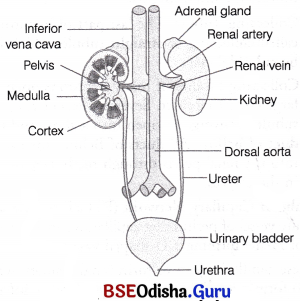
Human excretory system consists of a pair of kidneys,a pair of ureters, a urinary bladder and urethra, these are described below in detail
Human excretory system
Kidneys:
These are reddish brown, bean-shaped structures situated between the levels of last thoracic and third lumbar vertebra. These are close to the dorsal inner wall of the abdominal cavity.
Each kidney of an adult human measures, 10-12 cm in length, 5-7 cm in width, 2-3 cm in thickness with an average weight of 120-170 gm (i.e., 150 gm in males and about 135 gm in females).
Kidneys are mesodermal in origin. They develop from mesodermal nephrostomes or mesomeres, i.e. ciliated structures, functional in embryonic conditions.
Position of Kidneys:
The kidneys are located below the diaphragm on the left and right sides. The right kidney is lower and smaller than the left kidney because the liver takes up much space of the right side.
Structure of Kidney:
Structure of kidney can be studied well under two heads, i.e., external as well as internal structure.These are described below as
External Structure:
The outer surface of each kidney is convex. Inne/ concave surface has a notch called hilum. The supply of blood occurs through hilum. If we look from outside to inside, three layers cover the kidneys. These include renal fascia (outermost), the adipose layer and then renal capsule (innermost layer).
These coverings protect the kidneys from external shocks and injuries.
Internal Structure:
The LS of a mammalian kidney shows differentiation into outer cortex and inner medulla.
LS of kidney
Inside the kidney, the ureter is expanded as a funnel-shaped cavity called pelvis. The free end of pelvis has number of cup-like cavities called major calyces (sing, calyx) and minor calyces.
Medulla projects into the calyces as conical processes, called renal pyramids or medullary pyramids. The tip of pyramids are called renal papillae. The cortex spreads in between medullary pyramids as renal columns called columns of Bertini.
Microscopic Structure:
Each kidney is composed of numerous (nearly one million) complex tubular structures called nephrons. These are the functional units of kidney.
Functions of Kidney:
Following functions are served by kidney
(i) Regulation of water and electrolyte balance.
(ii) Regulation of arterial pressure.
(iii) Excretion of metabolic waste and foreign chemicals.
(iv) Secretion of hormones like renin, erythropoietin, etc.
Question 2.
Give an account of the structure and functions of the human kidney.
Answer:
Human excretory system consists of a pair of kidneys,a pair of ureters, a urinary bladder and urethra, these are described below in detail
Human excretory system
Kidneys:
These are reddish brown, bean-shaped structures situated between the levels of last thoracic and third lumbar vertebra. These are close to the dorsal inner wall of the abdominal cavity.
Each kidney of an adult human measures, 10-12 cm in length, 5-7 cm in width, 2-3 cm in thickness with an average weight of 120-170 gm (i.e., 150 gm in males and about 135 gm in females).
Kidneys are mesodermal in origin. They develop from mesodermal nephrostomes or mesomeres, i.e. ciliated structures, functional in embryonic conditions.
Position of Kidneys:
The kidneys are located below the diaphragm on the left and right sides. The right kidney is lower and smaller than the left kidney because the liver takes up much space of the right side.
Structure of Kidney:
Structure of kidney can be studied well under two heads, i.e., external as well as internal structure.These are described below as
External Structure:
The outer surface of each kidney is convex. Inne/ concave surface has a notch called hilum. The supply of blood occurs through hilum. If we look from outside to inside, three layers cover the kidneys. These include renal fascia (outermost), the adipose layer and then renal capsule (innermost layer).
These coverings protect the kidneys from external shocks and injuries.
Internal Structure:
The LS of a mammalian kidney shows differentiation into outer cortex and inner medulla.
LS of kidney
Inside the kidney, the ureter is expanded as a funnel-shaped cavity called pelvis. The free end of pelvis has number of cup-like cavities called major calyces (sing, calyx) and minor calyces.
Medulla projects into the calyces as conical processes, called renal pyramids or medullary pyramids. The tip of pyramids are called renal papillae. The cortex spreads in between medullary pyramids as renal columns called columns of Bertini.
Microscopic Structure:
Each kidney is composed of numerous (nearly one million) complex tubular structures called nephrons. These are the functional units of kidney.
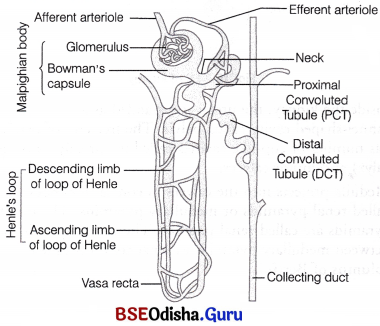
Structure of Nephron or Uriniferous Tubule:
Each nephron consists of two parts, i.e., the Malpighian body or renal corpuscle and the renal tubule.
(i) Malpighian Body or Renal Corpuscle Glomerulus along with Bowman’s capsule is called the Malpighian body or renal corpuscle which filters out large solutes from the blood and delivers small solutes to the renal tubule for modification.
Malpighian body (renal corpuscle)
Glomerulus is a tuft of thin-walled capillaries formed by the branching of afferent arteriole (a fine branch of renal artery).
Bowman’s Capsule (Glomerular capsule) It is a double-walled, cup-like structure that surrounds the glomerulus. The outer parietal wall of glomerulus is composed of flattened (squamous) cells and the inner visceral wall is composed of a special type of less flattened cells, called podocytes.
Diagrammatic representation of a nephron
(ii) Renal Tubules
Attached to each Bowman’s capsule is a long, thin tubule with three distinct regions.
These regions are described as follows
(a) Proximal Convoluted Tubule (PCT) Behind the neck, it makes few coils and is restricted to the cortical region of the kidney.
(b) Henle’s Loop It is quite narrower and U-shaped (or hair pin-shaped) having a descending limb that ends into the medulla and an ascending limb that extends back from the medulla into the cortex.
Differences between descending limb and ascending limb of henle’s loop
| Descending limb | Ascending limb |
| It is very thin. | It is thick. |
| Direction of fluid flow is downward. | Direction of fluid flow is upward. |
| Permeable to water. | Impermeable to water. |
(c) Distal Convoluted Tubule (DCT) is thicker and highly, coiled structure situated in the cortex like the proximal tubule. Coils of both convoluted tubules are intermingled.
(d) Collecting Tubule The last part of nephron is called collecting or straight tubule, which is lined by cuboidal cells.
Collecting tubules of many nephrons open into a large collecting duct. Several adjacent collecting tubules converge to open into a short and thick duct of bellini. All ducts of Bellini, then open into the renal pelvis through medullary pyramids in the calyces.
Peritubular Capillary Network (PTCN) is formed when a minute vessel of peritubular capillaries run parallel to the loop of Henle forming a U-shaped vasa recta.
All these capillaries join to form renal venules, which join to form a renal vein that opens into the inferior vena cava.
Types of Nephrons:
Based on the location in the kidney, nephrons are of following two types
(i) Cortical Nephrons:
In majority of nephrons, the loop of Henle is too short and extends only very little into the medulla, i.e., lie in the renal cortex. Such nephrons are called cortical nephrons.
(ii) Juxtamedullary Nephrons:
In some of the nephrons, the loop of Henle is very long and runs deep into the medulla. These nephrons are called juxtamedullary nephrons.
Note The cortical nephron forms about 80% of the total nephron count while rest 20% are the juxtamedullary nephron
Blood Supply to the Nephron:
Blood to each kidney is supplied by a renal artery and is drained by the renal vein. Both these blood vessels pass through hilus. The renal artery after entering into kidney divide into afferent arterioles each afferent arteriol form a tuft of capillaries in glomerulus. The capillaries reunite to form the efferent arteriole.
The afferent arteriole is short and wide that supplies blood to the glomerulus, while, the efferent arteriole is narrow and long carrying blood away from the glomerulus.
Differences between afferent arteriole and efferent arteriole
| Afferent arteriole | Efferent arteriole |
| These are the fine branches of renal artery which enter in each kidney and arise from dorsal aorta. | These are the fine branches of renal vein which leave the kidney and joins with inferior vena cava. |
| They bring arterial blood to the renal corpuscles. | They carry venous blood away from the renal corpuscles. |
| Its diameter is more. | Its diameter is comparatively less. |
| The blood flowing in it is rich in waste products. | The blood flowing in it is poor in waste products. |
Functions of Kidney:
Following functions are served by kidney
(i) Regulation of water and electrolyte balance.
(ii) Regulation of arterial pressure.
(iii) Excretion of metabolic waste and foreign chemicals.
(iv) Secretion of hormones like renin, erythropoietin, etc.
Question 3.
Give an account of the mechanism of urine formation.
Answer:
Formation of Urine:
The formation of urine is the result of processes like glomerular filtration, selective reabsorption and tubular secretion. These processes occur in different parts of nephrons. Details of these are as follows
Ultrafiltration or Glomerular Filtration:
The first step of urine formation is the filtration of blood. It is carried out by the glomerulus. That is why this step is called glomerular filtration.
Kidneys filter about 1100-1200 mL of blood per minute, which constitute roughly 1/5th of the blood pumped out by each ventricle of the heart in a minute.
The glomerular capillary blood pressure causes filtration of blood through three layers, i.e.,
(i) the endothelium of glomerular blood vessels.
(ii) the epithelium of Bowman’s capsule.
(iii) a basement membrane (present between the above mentioned two layers).
The podocytes (epithelial cells of Bowman’s capsule) are arranged in such a manner, so that some minute spaces called filtration slits or slit pores occur between them.
Diagram representing the path taken by fluid (glomerular filtrate) during its passage from the plasma in a glomerular capillary of the lumen of a renal capsule
On account of the high pressure in the glomerular capillaries, the substances are filtered through these pores into the lumen of the Bowman’s capsule (but the RBC, WBC and plasma proteins having high molecular weight unable to pass out).
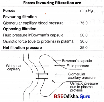
Varying pressures that operates between glomerular capillary and the Bowman’s capsule.
That’s why this process of filtration through glomerular capillaries in the Bowman’s capsule is known as ultra filtration and the filtrate is called glomerular filtrate.
It is hypotonic to urine that is actually excreted. Basic function of nephron is to clear out the plasma from unwanted substrates and also maintain the osmotic concentration of the blood plasma. Thus, the fluid coming out is known as urine, whose formation occurs inside the kidney.
Glomerular Filtration Rate:
The amount of the filtrate formed by the kidneys per minute is called Glomerular Filtration Rate (GFR). In a healthy person it was found approximately 125 mL /min, i. e., 180 L/day. GFR is regulated by one of the efficient mechanism carried out by Juxtaglomerular Apparatus (JGA).
JGA is a special sensitive region formed by cellular modifications in the distal convoluted tubule and the afferent arteriole at the location of their contact.
This apparatus includes
(i) granular juxtaglomerular cells in the afferent arteriole.
(ii) macula densa cells of DCT.
(iii) agranular lacis cells situated in between the above two.
Question 4.
Give an account of the role of the human kidneys in osmoregulation.
Answer:
Osmoregulation refers to the control of water level in the body. When there is excess of water in the body, kidney produces large volume of dilute urine and brings the water level to correct position. In the event of deficiency of water, kidney helps to conserve body water by producing small.