
Odisha State Board CHSE Odisha Class 11 Biology Solutions Chapter 6 Anatomy of Flowering Plants Textbook Questions and Answers.
CHSE Odisha 11th Class Biology Chapter 6 Question Answer Anatomy of Flowering Plants
Anatomy of Flowering Plants Class 11 Questions and Answers CHSE Odisha
Very Short Answer Type Questions
Fill in the blanks
Question 1.
Companion cells are associated with …………… .
Answer:
Sieve elements
Question 2.
Numerous vascular bundles are scattered in the ground tissue of …………. stem.
Answer:
Monocot
Question 3.
Vascular bundles in which phloem is found on both sides of xylem are called …………. .
Answer:
Bicollateral
Question 4.
The inner, darker and harder portion of secondary xylem that cannot conduct water in an older stem is called ………. wood.
Answer:
Heart
Write True or False. Then correct the sentences without changing the underlinked worlds
Question 1.
Conjoint vascular bundles are those in which xylem and phloem occur in one strand.
Answer:
True
Question 2.
In maize stem bicollateral vascular bundles are seen.
Answer:
In Cucurbita stem bicollateral vascular bundles are seen.
Question 3.
Vascular tissue system develops from ground meristem.
Answer:
Vascular tissue system develops from procambium.
Question 4.
Epidermal tissue system originates from protoderm.
Answer:
True
Question 5.
Endodermis is the outer Layer of stele.
Answer:
Pericycle is the outer Layer of stele.
Question 6.
Outer layer of tunica always gives rise to epidermis.
Answer:
True
Question 7.
Bulliform cells belong to epidermal tissue system.
Answer:
True
Question 8.
Epistomatic leaves are generally found in hydrophytes.
Answer:
True
Question 9.
Casparian strips are seen in pericycle layer.
Answer:
Casparian strips are seen in hypodermis.
Question 10.
Starch sheath refers to cells of pericycle with starch.
Answer:
Starch sheath refers to cells of endodermis with starch grains.
Short Answer Type Questions
Differentiate between the following
Question 1.
Spongy parenchyma and palisade parenchyma
Answer:
Differences between palisade and spongy parenchyma are
| Palisade Parenchyma | Spongy Parenchyma |
| These are present below the upper epidermis in dorsiventral leaf. | These are present towards the lower epidermis in dorsiventral leaf. |
| These are vertically elongated parenchymatous cells and are tightly fitted to each other without intercellular spaces. | These are oval-shaped parenchymatous cells and are loosely arranged with large air chambers. |
| These are present in three layers. | These are present in multilayers. |
| These cells contain more chloroplasts, which are radially arranged in these cells. Thus, the upper part of the leaf is dark green. | These cells contain few chloroplasts, which are irregularly distributed. Thus, lower part of the leaf is less green. |
Question 2.
Vascular bundles of Maize and Tridax
Answer:
Differences between vascular bundles of maize and Tridax are
| Vascular bundles of Maize | Vascular Bundles of Tridax |
| Maize is a monocot. | Tridax is a dicot |
| In stem | In stem |
| Vascular bundles are scattered in the ground tissue system. | Vascular bundles are arranged in a ring. |
| Vascular bundles are comparatively more in number. | Vascular bundles are fewer in number. |
| Vascular bundles are elliptical or rounded. | Vascular bundle are wedge shaped. |
| Vascular bundles are open. | Vascular bundles are closed. |
| Vascular bundle are not surrounded by any bundle sheath. | Vascular bundles are |
| In Root | In Root |
| Vascular bundles are polyarch and radial. | Vascular bundles are four in number arranged on alternate radii. |
Question 3.
Heartwood and sap wood.
Answer:
Differences between sapwood and heartwood are
| Sapwood | Heartwood |
| The outer region of the old trees forms the sap wood. | The central region of the old trees forms the heart wood. |
| It is also called as alburnum. | It is also called as duramen. |
| It is soft and not durable. | It is hard and durable. |
| It is light coloured and formed of living cells. | It is dark coloured due to the deposition of various substances. |
| Vessels are not blocked by tyloses. | Vessels are blocked by tyloses with various deposits. |
| The function of this region is conduction of water and nutrients and also storage of food. | The function of this region is mechanical support. |
Question 4.
Permanent tissue and meristematic tissue.
Answer:
Differences between meristematic and permanent tissues
| Meristematic tissue | Permanent tissue |
| Continuously dividing cells. | Do not divide continuously |
| Cells are small and isodiametric | Variable in shape and size |
| Cell wall is thin | Cell wall is thick |
| Nuclei are large | Nuclei are small |
| Vacuoles are absent | Vacuoles are present |
| Intercellular spaces absent | Intercellular spaces present |
| Metabolic activities are at high rate | Low rate |
| Inorganic inclusion absent | Present |
| Cell are undifferentiated | Cells are differentiated |
| Simple tissue | Simple or complex |
Question 5.
Spring wood and autumn wood.
Answer:
Differences between spring wood and autumn wood are
| Spring wood (Early wood) | Autumn wood (Late wood) |
| 1. Wood formed in favourable season is called spring wood. | Wood formed in unfavourable season is called autumn wood. |
| 2. The band of xylem is broad. | The band of Xylem is narrow. |
| 3. Xylem has wider lumen and thin walled vessels. | Xylem has narrower lumen with thick walled vessels |
| 4. Lighter in colour. | Comparatively darker |
| 5. Fibres are less in number | Fibers are abundant. |
Question 6.
Cork and bark.
Answer:
Differences between cork and bark are
| Cork | Bark |
| 1. Cork is a part of bark arising through the division of cork cambium. | Bark is the protective; outer layer of a woody tree. |
| 2. Cork is formed through cork cambium. | Bark is formed through both cork and vascular cambium. Bark consists of the cork, cork cambium and the secondary phloem. |
| 3. Cork consists of dead cells, which are filled with suberin. | Bark contains live tissues like cork cambium and secondary phloem. |
Write note on
Question 1.
Cambium
Answer:
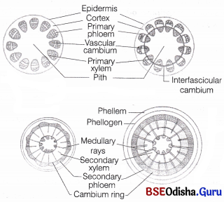
Formation of Cambium Ring
The parenchyma cells of the primary medullary rays adjacent to the intrafascicular cambium undergo dedifferentiadon and give rise to interfascicular cambium.
This joins the intrafascicular cambium of either side to form a complete ring of meristem called the cambium ring.
Question 2.
Lateral meristem
Answer:
Lateral Meristems:
The meristems that occur in the mature regions of roots and shoots of many plants. These meristems produce woody axis. These appear later than primary meristem, so are also called the secondary meristem.
They are cylindrical meristems. Some examples of lateral meristems are fascicular vascular cambium, interfascicular cambium and cork cambium. These are responsible for producing the secondary tissues.
Question 3.
Bark
Answer:
Bark:
Bark is a non-technical term used to describe all tissues exterior to the vascular cambium, therefore including secondary phloem. The bark refers to a number of tissues, i.e., periderm and secondary phloem. The bark that is formed early in the season is called early or soft bark. Towards the end of the season, late or hard bark is formed.
Question 4.
Lenticels
Answer:
Lenticels:
At certain regions of stem, the phellogen cuts off closely arranged parenchymatous cells on the outer side instead of cork cells.
These parenchymatous cells soon rupture the epidermis forming a lens-shaped opening called lenticels. The lenticels are mostly found in woody trees.
Question 5.
Hydathode
Answer:
These are specialised pores along the margins and apex of the leaf through which the secretion of water (guttation) takes place. Hydathodes or water stomata consists of vein endings, epithem chamber and pores. Pores are surrounded by guard cells, but remains open permanently.
Question 6.
Promeristem
Answer:
Procambium:
Ir develops into primary vascular tissue. It forms the isolated strands of elongated cells, very near to the central region.
Question 7.
Glandular tissues.
Answer:
Glandular Tissues These have unicellular or multicellular glands, which may secrete or excrete chemicals. They are present either externally or internally.
- External glands are superficial in nature and formed on epidermis. They may occur as hydathodes glandular hairs, nectaries and digestive glands, etc.
- Internal glands are confined to internal tissues.
They are formed by lysis of some of the cells or by splitting of cells at middle lamella. They occur as oil glands, mucilage secreting glands, etc.
Question 8.
Annual ring.
Answer:
In tropical areas, the growth of secondary xylem is continuous. In others, yearly growth is very distinct and appears in the form of annual rings.
The transition from springwood to autumnwood is gradual. After autumnwood and before springwood of next year, there is no growth.
Therefore, change over from autumnwood to springwood is sudden.
The light coloured springwood and its next dark coloured autumnwood constitutes an annual ring or growth ring. It represents the total secondary xylem or wood formed in one year.
Question 9.
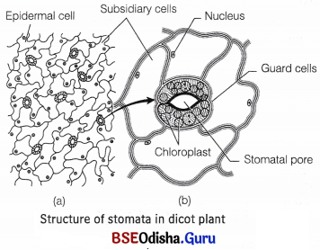
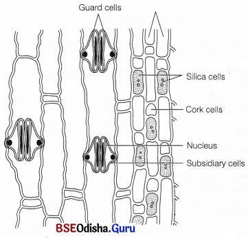
Different types of stomata
Answer:
The stomata (sing, stoma) are openings in the epidermis of most of the aerial parts of the plants, especially the leaves. Each stomata is composed of two bean-shaped cells called as guard cells, which enclose stomatal pore. The guard cells are generally much smaller in size as compared to other epidermal cells.
They are sensitive to even a small change in turgor pressure. The dimension of stomatal pore varies from species to species, but it measures about 20 Jim in length and about 10-20 jlm in width when fully open.
In some species, the guard cells are surrounded by subsidiary cells or accessory cells which differ morphologically from the other epidermal cells. The guard cell walls have special elastic properties. The adjoining cell walls of two guard cells around pore are free and not attached with each other.
These properties help them to stretch laterally during stomatal opening. The stomatal aperture, guard cells and the surrounding subsidiary cells are together called stomatal apparatus.
In most monocots, the guard cells are dumb-bell-shaped. The stomata are mostly found on the upper epidermis of the leaves. In some hydrophytes, the stomata occur on the upper surface to avoid water contact.
Question 10.
Trichomes
Answer:
Trichomes The epidcrmal hairs present on the stem are called trichomes. These are epidermal outgrowths present temporarily or permanently on almost all plant parts. They are multicellular structures which may he branched or unbranched. They may be secretory in nature.
They also help in checking the rate of transpiration from aerial plant surfaces.
Long Answer Type Questions
Question 1.
Compare the internal structure of a dicot stem with that of a dicot root.
Answer:
Dicotyledonous Stem:
The transverse section (TS) of a typical young dicotyledonous stem (e.g. Halianthus annus, Cucurcubita maxima, etc.) shows the following areas
Epidermis:
The outermost protective layer of the stem is called epidermis. It is covered with a thin-layer of cudcle and may bear trichomes and a few stomata.
The cuticle protects the tissues from injury as well as diseases from the entry of fungal spores and bacteria. It also helps to prevent loss of water.
Epidermal Epidermis Hypodermis Parenchyma Endodermis Pericycle Vascular bundle Medullary rays
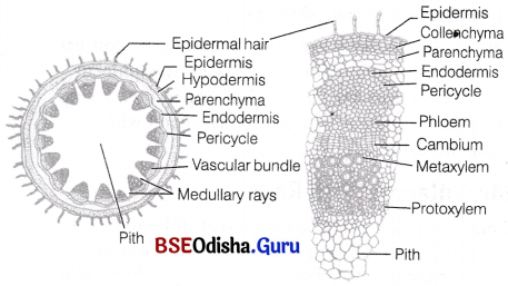
Cortex:
This layer lies just below the epidermis and extends till ” endodermis. Its various parts are hypodermis, general cortex and endodermis.
(i) Hypodermis
It is just below the epidermis consisting of collenchymatous cells. The cells contain chloroplasts. It provides mechanical strength to the stem.
(ii) General Cortex
It is located just below the hypodermis and consists of a few layers of parenchymatous cells. These cells are thin-walled have big intercellular spaces and may contain chloroplasts.
(iii) Endodermis
It lies just beneath the general cortex in the form of single layer of barrel-shaped cells surrounding the stele. It is the innermost layer of cortex. In sunflower, it contains starch, hence is called starch sheath.
Dicotyledonous Root:
The primary internal structure of dicot root (e.g. gram) can be studied from the Transverse Section (TS) of a young root of sunflower, pea or gram. The primary root is the one which has only primary permanent tissues that are formed from vegetative shoot apex. Secondary tissues are absent.
The following structures can he seen from periphery towards the centre
Epidermis:
It forms the outermost layer in young root. It is equivalent to epidermis of stem. The stomata and cuticle are not present in it. The cells are thin-walled and tubular. Some of the epidermal cells are prolonged to form thin-walled tubular structures called root hairs. The cells which produce root hair are called root hair cells or trichoblasts. Due to the presence of root hairs, epiblema is also called piliferous layer (Pilus – hair; ferre – to carry) and rhizodermis (Rhiza – root; derma – skin).
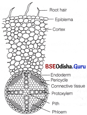
TS of a typical dicot root
Root hairs have pectose layer on the outside, this is to help them to pass into the soil spaces for absorption of water and mineral salts. The active lifespan of root hairs is upto 7 days and they die off in older parts of the root.
The cells of older epidermis shrivel afterwards and become cutinised and suberised.
Cortex:
It lies beneath the epiblema. It consists of several layers of thin-walled parenchymatous cells with conspicuous intercellular spaces. The cells of cortex store food. It also conducts water from the epiblema to the inner tissues.
Endodermis:
The innermost layer of the cortex is endodermis. It comprises of a single layer of barrel-shaped cells without any intercellular spaces. The endodermal cells are living and are rich in starch grains.
They have characteristic bands of thickenings along their radial and tangential walls. These are called casparian bands or casparian strips.
The casparian strips are made up of suberin and lignin. These strips prevent plasmolysis of endodermal cells and do not allow wall to wall movement of substances, between cortex and pericycle.
The cells of endodermis lying opposite to the protoxylem are thin-walled to permit free passage of water and minerals from cortex into the xylem. These are called passage cells.
Question 2.
Describe the anatomy of a typical monocot stem with labelled diagram. Point out the differences in the structure of vascular bundles in monocot stem and dicot stem.
Answer:
Monocotyledonous Stem:
The monocot stem (e.g. Zea mays, Canna, etc.) possesses only primary structure. The different monocot stems from outside towards inside consist of epidermis, hypodermis, ground tissue and vascular system.
Epidermis:
It is single-layered, having stomata in it. The cells have a thick cuticle layer on the outside.
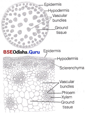
TS of monocot stem
Hypodermis
It is 2-3 layered having lignified scierenchymatous cells present just below the epidermis.
Ground Tissue
It fills the whole interior of the stem containing parenchymatous cells. A number of vascular bundles are scattered in it.
Vascular System
Monocot stems has scattered vascular bundles.
Each vascular bundle in vascular strand is surrounded by a sheath of sclerenchyma known as bundle sheath cells. The vascular bundles possess both phloem (phloem parenchyma is absent) and xylem, so these are conjoint type.
The bundles are endarch with the protoxylem and metaxylem are arranged in the form of a ‘Y’. The divergent ends are occupied by two pitted vessels and convergent end by two smaller spiral vessels lying radially in the centre. A water containing cavity called lysigenous cavity is present in association with the protoxylem.
It is formed by the breakdown of inner protoxylem vessels and parenchyma during the earlier stages of growth. The cavity is absent or reduced in the smaller vascular bundles that occur in contact with sclerenchymatous hypodermis.
A brief account of internal structure of Zea mays and Canna has been described here under.
Differences between vascular bundles in monocot stem
| Vascular Bundle in Monocot Stem | Vascular Bundle in Dicot Stem |
| Vascular bundles are arranged in a ring. This is called stelar region. | Vascular bundles are scattered in the ground tissue system. |
| Vascular bundles are fewer in number. | Vascular bundles are comparatively more in number. |
| Vascular bundles are wedge-shaped. | Vascular bundles are elliptical or rounded. |
| Vascular bundles are surrounded by bundle sheath | Vascular bundles are not surrounded by any bundle sheath. |
| Vascular bundles are closed | Vascular bundles are open. |